Keith D. Sudheimer, Brian M. Winn, Jay M. Shoaps, Kristina K. Davis, Archibald J. Fobbs Jr., and John I. Johnson
Radiology Department, Communications Technology Laboratory, and College of Human Medicine, Michigan State University;
National Museum of Health and Medicine, Armed Forces Institute of Pathology
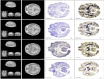
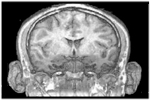
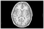
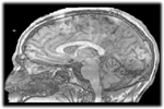
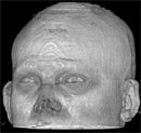
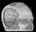
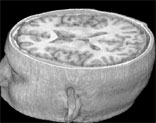
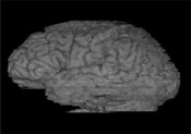
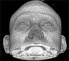
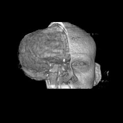
In this atlas you can view MRI sections through a living human brain as well as corresponding sections stained for cell bodies or for nerve fibers. The stained sections are from a different brain than the one which was scanned for the MRI images. Furthermore, for the stained sections, the brain was removed from the skull, dehydrated, embedded in celloidin, cut with a sliding microtome, passed through several staining and differentiating solutions, and mounted on glass slides. Each step of these procedures changed the shaped of the brain and of the sections. Therefore the stained sections will be quite a different size and shape than those of the MRI sections. Nevertheless, comparing MRI images with stained sections from approximately the same level can greatly increase understanding of the internal architecture of these brains.
Please use the images and data from this site.
Click here for instructions
|
|
|||
|
Coronal Plane
|
Horizontal Plane
|
Sagittal Plane
|
|
 |
|||
|
Or View Movies Of Every Section Through the Brain
|
|||
|
(Movie - 4.2MB) |
Horizontal MRI Movie
(Movie - 2.5MB) |
(Movie - 2.8MB) |
|
Or click on a picture below to see a movie:
|
(VR object - 3.0MB) |
(Movie - 2.7MB) |
(Movie - 3.9MB) |
|
All Views |
Just Brain |
Nose |
Half Brain |
Download The Human Brain Screensaver for PC 12MB
See also:
Supported by: Grants 9812712, 9814911, and 9814912, from the National Science Foundation
Division of Integrative Biology and Neuroscience.
