John I. Johnson, Brian M. Winn, Joseph J. Maleszewski, Myrvine Bernadotte, Prashant Vaishnava and Keith D. Sudheimer
Radiology Department, Communications Technology Laboratory, and College of Human Medicine, Michigan State University;
return to main menu
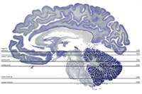
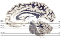
In this atlas you can view axial sections stained for cell bodies or for nerve fibers, at six rostro-caudal levels of the human brain stem.
Please use the images and data from this site.
Click here for instructions
Click Below to Select Levels and Sections to View.

Cell Selection and Level Selector
|
|

Fiber Selection and Level Selector
|
Supported by: Grants 9812712, 9814911, 9814912, 0131028, 0131267, 0131826 from the National Science Foundation
Division of Integrative Biology and Neuroscience.
Images from the Yakovlev-Haleem brain collection are used courtesy of the National Museum of Health and Medicine, Armed Forces Institute of Pathology,
Adrianne Noe, Ph. D., Director; Archie J. Fobbs, Jr., Neuroanatomical Curator.

