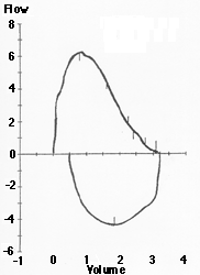
Normal Flow Volume Loop
Interpretation of Pulmonary Function Tests: Spirometry: Interpreting Spirometry
Thomas Gross, MD
Peer Review Status: Externally Peer Reviewed by the Department of Internal Medicine Virtual Hospital Editorial Board
Normal values for FEV1 and FVC are based on population studies and vary according to race, height, age, and gender. They are expressed in both absolute numbers and percent predicted of normal. Some authors have suggested that defining normal by 95% confidence intervals would be more statistically appropriate, particularly at the extremes of age. Thus, a value below the 5th percentile is defined as "below the lower limit of normal." However, many laboratory and computer software programs continue to express results as percentages of predicted normal values. A physician's interpretative strategy should be adaptable to either reporting system.
Values for FVC and FEV1 that are over 80% of predicted are defined as within the normal range. The FEV1/FVC ratio is expressed as a percentage, and a normal young individual is able to forcibly expire at least 80% of his/her vital capacity in one second. A ratio under 70% suggests underlying obstructive physiology; however, the FEV1/FVC ratio declines as a normal sequelae of aging. Thus, at advanced ages, pathologic airways obstruction is diagnosed based upon deviation from predicted FEV1/FVC values, with values below the 5th percentile best selecting patients with obstructive defects.
|
Normal Flow Volume Loop
|
A normal flow volume loop has a rapid peak expiratory flow rate with a gradual decline in flow back to zero. The inspiratory portion of the loop is a deep curve plotted on the negative portion of the flow axis. Inspiratory data is often overlooked, but the flow volume loop gives the opportunity of assessing this information as well.
|
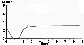
Normal Volume Time Curve
|
The normal volume time curve has a rapid upslope and approaches a plateau soon after exhalation. The maximum volume attained represents the forced vital capacity (FVC), while the volume attained after one second represents the forced expiratory volume (FEV1).