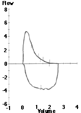
Mild Obstruction Flow Volume
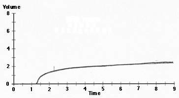
Mild Obstruction Volume Time Curve
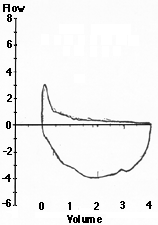
Severe Obstruction Flow Volume
Interpretation of Pulmonary Function Tests: Spirometry: Interpreting Spirometry
Thomas Gross, MD
Peer Review Status: Externally Peer Reviewed by the Department of Internal Medicine Virtual Hospital Editorial Board
|
FEV1 |
> 80% predicted |
normal |
|
65 - 80% |
mild |
|
|
50 - 65% |
moderate |
|
|
< 50% |
severe |
As the obstruction becomes more severe and end expiratory air trapping develops, the forced vital capacity may be reduced as well as the FEV1; however there should continue to be a disproportionate reduction in FEV1 as evidenced by the FEV1/FVC ratio.
Example of spirometry results demonstrating mild obstruction:
|
Meas |
Pred |
%Pred |
|
|
FVC |
2.63 |
3.11 |
84 |
|
FEV1 |
1.58 |
2.28 |
69 |
|
FEV1/FVC |
60 |
73 |
|
|
FEF25-75 |
0.59 |
2.56 |
23 |
|
PEF |
4.90 |
5.78 |
85 |
|
Mild Obstruction Flow Volume
|
Mild Obstruction Volume Time Curve
|
|
Severe Obstruction Flow Volume
|
Obstructive lung disease also changes the appearance of the flow volume curve. As with a normal curve, there is a rapid peak expiratory flow, but the curve descends more quickly than normal and takes on a concave shape, reflected by a marked decrease in the FEF25-75. With more severe disease, the peak becomes sharper and the expiratory flow rate drops precipitously. This results from dynamic airway collapse which occurs as diseased conducting airways are more readily compressed during forced expiratory efforts. On the volume time curve, this is seen as a slower ascent to maximum volume, with a gradual upsloping versus the rapid rate seen in normal individuals. This equates with a prolonged forced expiratory time demonstrable on physical exam.
The ATS recommends caution in diagnosing obstruction when a patient has a reduced FEV1/FVC ratio but normal FEV1 and FVC. As mentioned above, there is a normal age-related decline in the FEV1/FVC ratio, so normal elderly patients without airway obstruction will have a ratio below 70-80%. In this case, values below the predicted FEV1/FVC ratio aid in diagnosing obstruction. The mid-range flows (FEF25-75) are always reduced in obstructive airways disease. However, some patients have normal spirometry with the exception of a reduced FEF25-75. While normal values for FEF25-75 have broader ranges than the other spirometirc values, a mid-range flow less than 50% is likely to be abnormal. This is suggestive of possible small airways dysfunction and potentially early obstruction, but it should not be interpreted as meeting obstructive criteria. In the appropriate clinical setting, one may consider a trial of bronchodilators, bronchoprovocative testing to exclude asthma, or interpret this observation as a possible early indicator of smoking related lung disease.