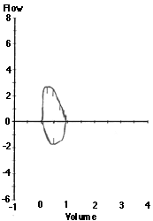
Restriction Flow Volume

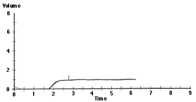
Restriction Volume Time

Interpretation of Pulmonary Function Tests: Spirometry: Interpreting Spirometry
Thomas Gross, MD
Peer Review Status: Externally Peer Reviewed by the Department of Internal Medicine Virtual Hospital Editorial Board
|
Meas |
Pred |
%Pred |
|
|
FVC |
0.96 |
2.75 |
35 |
|
FEV1 |
0.94 |
1.90 |
49 |
|
FEV1/FVC |
98 |
69 |
|
|
FEF25-75 |
2.25 |
2.11 |
107 |
|
PEF |
2.98 |
5.40 |
55 |
|
Restriction Flow Volume
|
Restriction Volume Time
|
The shape of the flow volume loop is relatively unaffected in restrictive disease, but the overall size of the curve will appear smaller when compared to normals on the same scale. Similarly, there will be a rapid upslope on the volume time curve, but such patients will reach a smaller vital capacity.
It is important to realize, however, that restrictive lung disease cannot be diagnosed by spirometry alone. With severe airways obstruction the lung volume remaining in the thorax after expiration (the functional residual capacity) may be increased to such a degree that it limits inspiratory capacity and, hence, reduces vital capacity. A reduced FEV1 and FVC are therefore only suggestive of true restrictive disease, but it is necessary to measure lung volumes to accurately diagnose restrictive physiology.